Back Of Neck Anatomy Muscles : Labeled Anatomy Chart Of Neck And Back Muscles On White ... - Intermediate back muscles and c.
byAdmin-
0
Back Of Neck Anatomy Muscles : Labeled Anatomy Chart Of Neck And Back Muscles On White ... - Intermediate back muscles and c.. Back muscles are divided into two specific groups: Related posts of muscle anatomy back of neck. There are many muscles around the neck that help to support the cervical spine and allow you to move your head in different directions. Sternohyoid, sternothyroid, thyrohyoid, omohyoid anterior vertebral muscles: Neck mobility is necessary primarily to rotate the head and keep the head upright.
As you know, the neck is the part of the body that sits between the head and torso. The back muscles stabilize and move the vertebral column, and are grouped according to the lengths and direction of the fascicles. There are several different layers of muscles in your back that are often pulling in different and the intermediate layer of back muscles includes the serratus posterior superior and inferior. C7 powers the triceps muscle on the back of your upper arms and transmits sensation along the back of the arms, and down to the middle finger. Muscles of the neck are described separately from the compartments.
Dental Gross Anatomy Lab 8 Posterior Triangle of the Neck ... from i.ytimg.com Muscles of the posterior neck and the back. As you know, the neck is the part of the body that sits between the head and torso. Spinous processes of txi to liii and supraspinous ligaments. The back muscles stabilize and move the vertebral column, and are grouped according to the lengths and direction of the fascicles. The pll starts at c2 and goes down the back of the vertebral bodies and intervertebral discs. Integrates anatomy and physiology of cells, tissues, organs, the systems of the human body, and mechanisms responsible for homeostasis. Extrinsic muscle layers of the back. The anterior and middle scalenes originate from the transverse processes of certain cervical vertebrae and attach to the first rib.
Alle muscles are detailed described incl.
The major muscle of the back of the neck, the trapezius, is involved in movements of the scapula and is dealt with in the next section, on the muscles in this view of a male figure with one arm up and one arm on the hip, there is a tremendous number of clearly defined anatomical shapes, large and small. Working in pairs on the left and. They work on the hyoid bone, with the suprahyoid muscles pulling up and the infrahyoid. Human muscle anatomy 12 photos of the human muscle anatomy human anatomy muscle questions, human anatomy muscles clay learning system, human muscle anatomy head, human muscle anatomy leg, human muscle. Neck flexion and homolateral tilt. Superficial muscles are the muscles closest to the skin surface and can usually be seen while a body is performing actions. This article describes the anatomy of the head and neck of the human body, including the brain, bones, muscles, blood vessels, nerves, glands, nose, mouth, teeth, tongue, and throat. As you know, the neck is the part of the body that sits between the head and torso. The neck muscles, including the sternocleidomastoid and the trapezius, are responsible for the gross motor movement in the muscular system of the head and neck. Sternohyoid, sternothyroid, thyrohyoid, omohyoid anterior vertebral muscles: The superficial group acts on upper limbs and. Digastric, mylohyoid, geniohyoid, stylohyoid infrahyoid muscles: Watch cervical muscle anatomy animation.
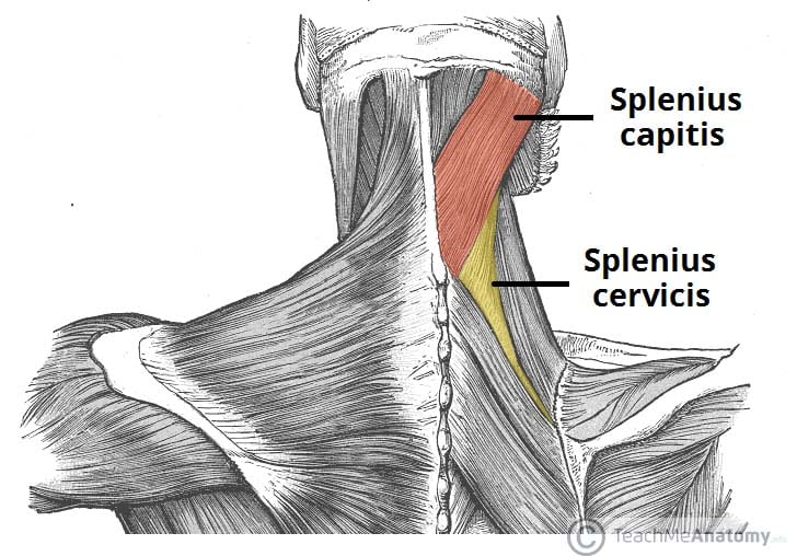
The posterior muscles of the neck are primarily concerned with head movements, like extension. Muscles of the neck are described separately from the compartments. The neck muscles, including the sternocleidomastoid and the trapezius, are responsible for the gross motor movement in the muscular system of the head and neck. Several other muscles of the back also extend up to the neck region and are partly connected with the cervical part of the vertebral column, including the trapezius, levator scapulae, splenius, iliocostalis, longissimus, rotatores, semispinalis, interspinales, and intertransversarii muscles. There are four pairs of muscles that are responsible for chewing movements or mastication.
How to Draw the Neck - Anatomy for Artists | Proko from www.proko.com Digastric, mylohyoid, geniohyoid, stylohyoid infrahyoid muscles: Superficial muscles are the muscles closest to the skin surface and can usually be seen while a body is performing actions. Several other muscles of the back also extend up to the neck region and are partly connected with the cervical part of the vertebral column, including the trapezius, levator scapulae, splenius, iliocostalis, longissimus, rotatores, semispinalis, interspinales, and intertransversarii muscles. The muscles of the back that work together to support the spine, help keep the body upright and allow twist and bend in many directions. The muscles of the back and neck that move the vertebral column are complex, overlapping, and can be divided into five groups. The neck has no external bone protective structures, so it is quite mobile. Human muscle anatomy 12 photos of the human muscle anatomy human anatomy muscle questions, human anatomy muscles clay learning system, human muscle anatomy head, human muscle anatomy leg, human muscle. The back muscles stabilize and move the vertebral column, and are grouped according to the lengths and direction of the fascicles.
The back anatomy includes the latissimus dorsi, trapezius, erector spinae, rhomboid, and the teres major.
The back muscles can be three types. Cervical spine anatomy is quite complex. There are four pairs of muscles that are responsible for chewing movements or mastication. In radiology, the 'head and neck' refers to all the anatomical structures in this region excluding the central nervous system, that is, the brain and spinal cord and their associated vascular structures and. Integrates anatomy and physiology of cells, tissues, organs, the systems of the human body, and mechanisms responsible for homeostasis. The superficial group acts on upper limbs and. There are several individual muscles within the back anatomy, and it's important to take a quick look the image below to shows all the major back muscles (as well as some neck muscles) They move the head in every direction, pulling the skull and jaw towards the shoulders, spine, and scapula. Neck flexion and homolateral tilt. This article describes the anatomy of the head and neck of the human body, including the brain, bones, muscles, blood vessels, nerves, glands, nose, mouth, teeth, tongue, and throat. The anterior and middle scalenes originate from the transverse processes of certain cervical vertebrae and attach to the first rib. We will attempt to provide a simplified overview of this complex anatomy. Human muscle anatomy 12 photos of the human muscle anatomy human anatomy muscle questions, human anatomy muscles clay learning system, human muscle anatomy head, human muscle anatomy leg, human muscle.
Cervical spine anatomy is quite complex. Understanding the anatomy of your cervical spine and the vital nerves it contains should motivate you to adopt behaviors that help prevent neck injury and. In anatomy, the neck is also called by its latin names, cervix or collum, although when used alone, in context, the word cervix more often refers to the uterine cervix, the neck of the uterus.3 thus the adjective cervical may refer. Related posts of muscle anatomy back of neck. Covers deep muscles of back and trunk.
The Intrinsic Back Muscles - Attachments - Actions ... from teachmeanatomy.info Intermediate back muscles and c. The muscles of the back and neck that move the vertebral column are complex, overlapping, and can be divided into five groups. They move the head in every direction, pulling the skull and jaw towards the shoulders, spine, and scapula. Several other muscles of the back also extend up to the neck region and are partly connected with the cervical part of the vertebral column, including the trapezius, levator scapulae, splenius, iliocostalis, longissimus, rotatores, semispinalis, interspinales, and intertransversarii muscles. Watch cervical muscle anatomy animation. Integrates anatomy and physiology of cells, tissues, organs, the systems of the human body, and mechanisms responsible for homeostasis. Sternohyoid, sternothyroid, thyrohyoid, omohyoid anterior vertebral muscles: Covers deep muscles of back and trunk.
The muscles of the back and neck that move the vertebral column are complex, overlapping, and can be divided into five groups.
The anatomy of your back muscles can be complex. These muscles course from your vertebral column to your ribs. Watch cervical muscle anatomy animation. The neck has no external bone protective structures, so it is quite mobile. This article describes the anatomy of the head and neck of the human body, including the brain, bones, muscles, blood vessels, nerves, glands, nose, mouth, teeth, tongue, and throat. This article covers the anatomy of the deep muscles of the back, including their function, blood supply, innervation, origin and insertion. The muscles of the back that work together to support the spine, help keep the body upright and allow twist and bend in many directions. Here the extrinsic back muscles are classified into logical subgroups to facilitate knowledge. There are several different layers of muscles in your back that are often pulling in different and the intermediate layer of back muscles includes the serratus posterior superior and inferior. Cervical spine anatomy is quite complex. As you know, the neck is the part of the body that sits between the head and torso. Related posts of muscle anatomy back of neck. Neck flexion and homolateral tilt.
Anterior muscles of the neck back of neck anatomy. Choose from 500 different sets of flashcards about anatomy back muscles neck thoracic on quizlet.